What’s more exciting than moving to a brand new lab space? Moving during a pandemic! The Computational and Systems Neuroscience Lab has officially (and safely) moved into the new science building at American University, the Hall of Science! Now all that’s left to do is finish setting up!
Kyra Swanson has successfully defended her dissertation (virtually!) on the Neural and Computational Mechanisms of Cognitive Flexibility. She’ll be continuing on at a position at the Smithsonian National Zoo to use deep learning and data analysis methods for ethological studies of zoo animals. Congratulations, Dr. Swanson!
Linda Amarante successfully defended her dissertation on the Distributed Processing of Reward Information by the Medial and Orbital Frontal Cortices. She’ll now be moving on to a post-doctoral position at Johns Hopkins University in Jeremiah Cohen’s lab. Congratulations Dr. Amarante!
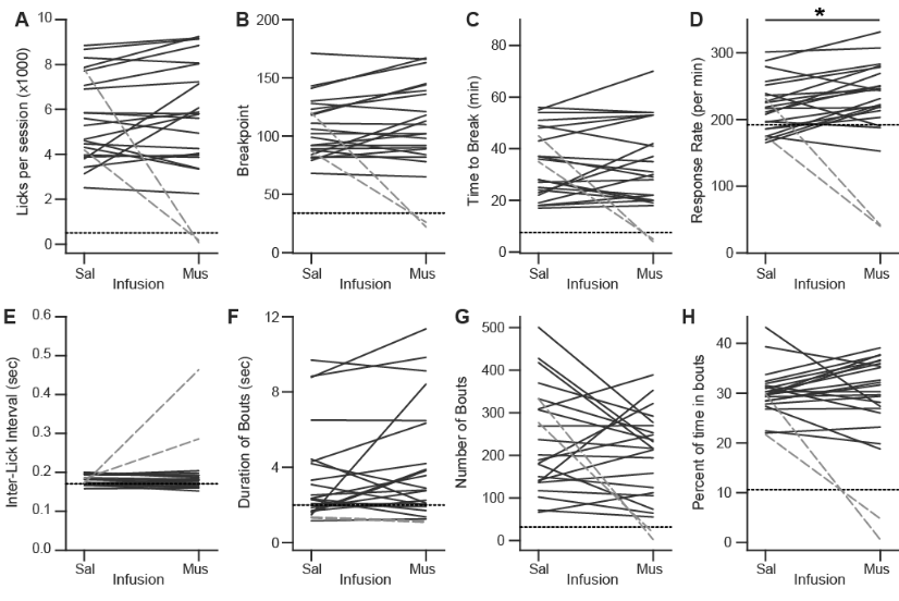
Check out our latest preprint turned publication in Behavioral Neuroscience!
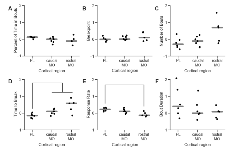
We used a progressive ratio licking task in which rats had to make increasing numbers of licks to receive liquid sucrose rewards. We determined what measures of progressive ratio performance are sensitive to value by testing rats with rewards containing 0%–16% sucrose. We found some measures (breakpoint, number of licking bouts) were sensitive to sucrose concentration and others (response rate, duration of licking bouts) were not. Inactivation of MFC had no effects on measures associated with value (e.g., breakpoint), but did alter behavioral measures associated with the pace of task performance (response rate and time to break). Our findings suggest that the medial frontal cortex has a role in maintaining task engagement, but not in the motivational control of action, in the progressive ratio licking task.
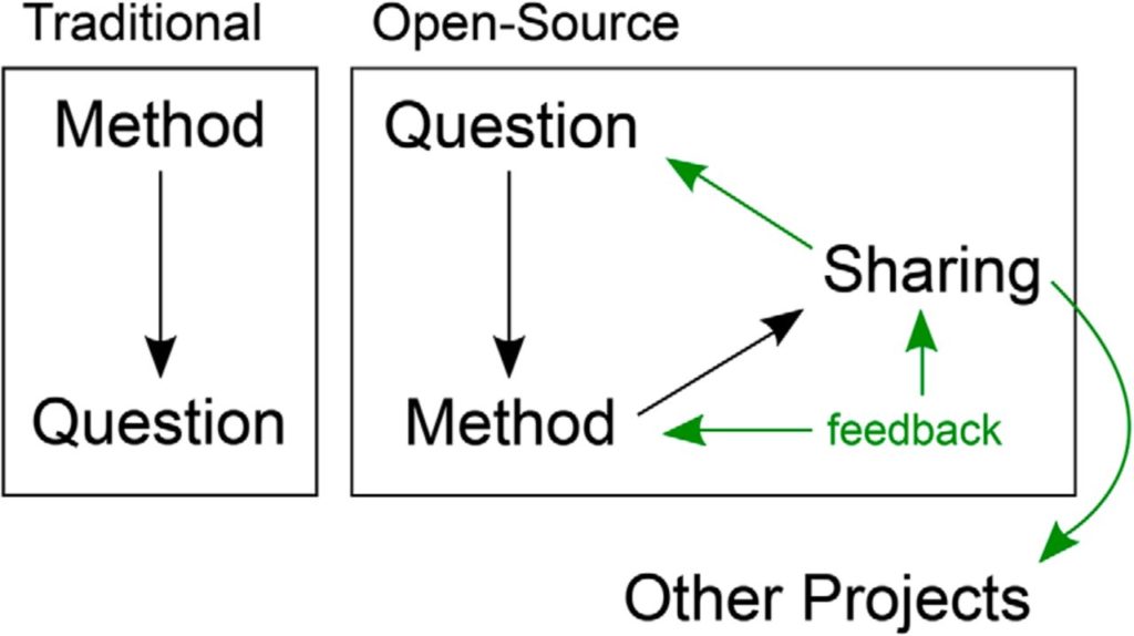
We just published a new commentary on the future of open-source tools for behavioral neuroscience, inspired by our work with OpenBehavior.com!
There has been a recent and substantial increase in the use of open-source tools for conducting research studies in neuroscience. The OpenBehavior Project was created to disseminate open-source projects specific to the study of behavior. In this commentary, we emphasize the benefits of adopting an open-source mindset and highlight current methods and projects that give promise for open-source tools to drive advancement of behavioral measurement and ultimately understanding the neural basis of behavior.
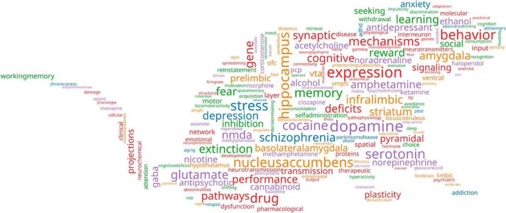
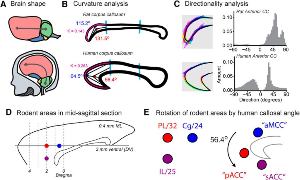
Our latest review, What, if anything, is rodent prefrontal cortex? is now published in eNeuro!
Studies on prefrontal parts of the rodent cerebral cortex have appeared at an increasing rate in recent years. However, there has been no consensus on the terms used to describe the rodent prefrontal cortex (PFC) or how it relates to the PFC of monkeys and humans. To address these issues, we conducted a meta-analysis of publications on the PFC across species, a review of rodent brain atlases, a survey of PFC researchers on anatomic terms, and an analysis of how species differences in the corpus callosum might help relate PFC areas across species. Addressing these issues may help improve the clarity, rigor, and reproducibility of research on the rodent PFC.
We’ve just posted our second preprint! This time, we wrote a review article discussing the complicated history, and hopefully clear future, of rodent prefrontal cortex.
Very exciting news! Our first preprint has made it to its final published form in the Journal of Neuroscience.
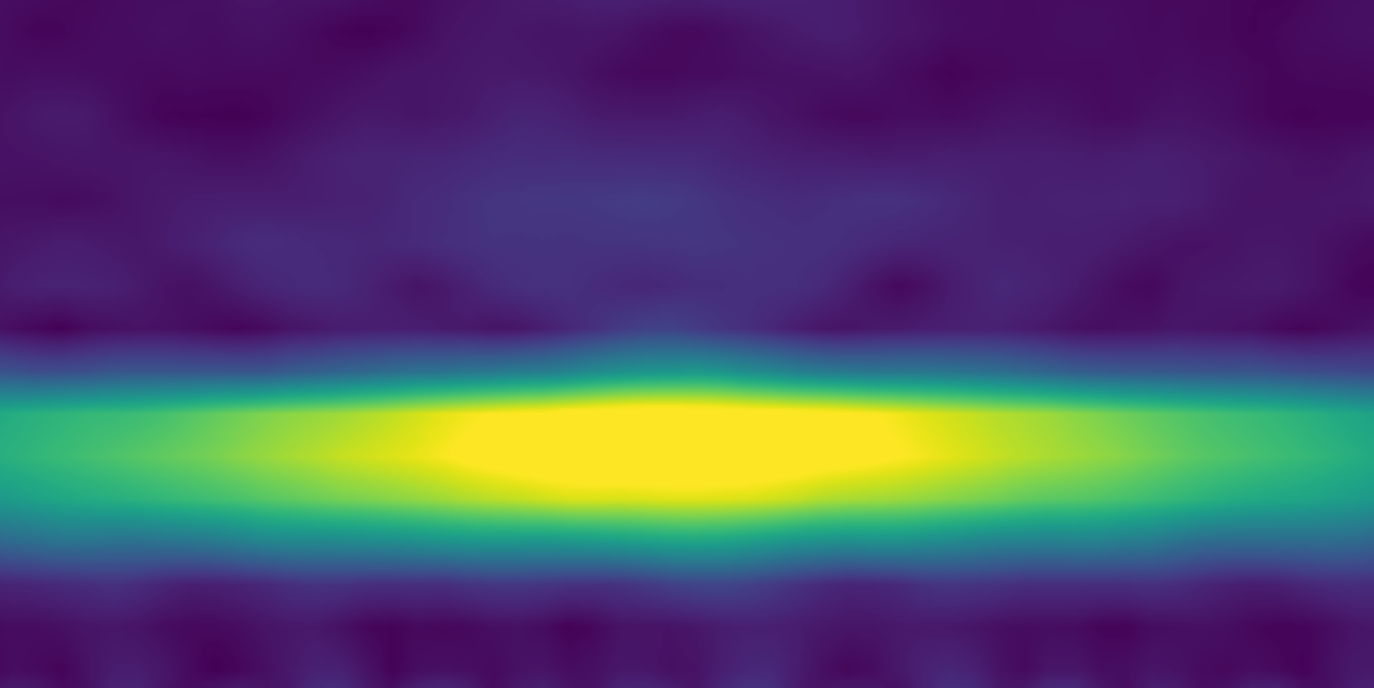
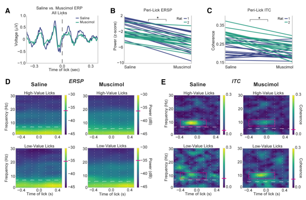
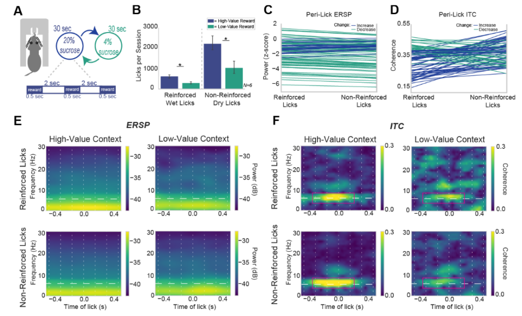
In our new work, we report evidence for a 6–12 Hz theta rhythm that is generated by the medial frontal cortex (MFC) and synchronized with ongoing consummatory actions. Previous studies of MFC reward signaling have inferred value coding upon temporally sustained activity during the period of reward consumption. Our findings suggest that MFC activity is temporally sustained due to the consumption of the rewarding fluids, and not necessarily the abstract properties of the rewarding fluid. Two other major findings were that the MFC reward signals persist beyond the period of fluid delivery and are generated by neurons within the MFC.
How do we know the reward value of a given food or fluid? The item must first be consumed and only then can its relative value be computed. Here, we investigated the relationship between licking and reward signaling by the medial frontal cortex (MFC), a key cortical region for reward-guided learning and decision-making. Rats were tested in an incentive contrast procedure, in which they received alternating access to higher and lower value sucrose rewards. Neuronal activity in the MFC encoded the relative value of the ingested fluids, showing stronger entrainment to the lick cycle when animals ingested higher value rewards. The signals developed with experience, encoded the reward context, and depended on neuronal processing within the MFC. These findings suggest that consummatory behavior drives reward signaling in the MFC.