Tag: 3D Print

3D Printed Headcap and Microdrive

SEPTEMBER 26, 2019

In their 2015 Journal of Neurophysiology article, the Paré Lab at the Center for Molecular and Behavioral Neuroscience at Rutgers University describe their novel head-cap and microdrive design for chronic multi-electrode recordings in rats through the use of 3D printing technology and highlight the impact of 3D printing technology on neurophysiology:


There is a need for microdrives and head-caps that can accommodate different recording configurations. Many investigators implant multiple individual drives aiming to record from numerous areas. However, this extends surgery time, impairs animal recovery, and complicates experiments. Other strategies rely on more expensive custom-machined drive assemblies that are specifically built for a particular set of regions, limiting their adaptability. Some proposed designs allow targeting of multiple regions, but recording sites must be within a few millimeters so are only suitable for mice and not for accessing areas of larger brains (like in rats, for example).

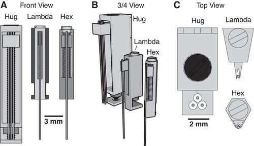
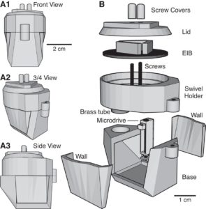
Utilizing 3D printing technology to create a novel design concept of microdrives and head-caps, this group’s design allows for recording of multiple brain regions in different configurations. In their article, the lab reviews the basic principles of 3D design and printing and introduce their approach to multisite recording, explaining how to construct the individual required components. The 3D printed head cap and electrode microdrive enables investigators to perform chronic multi-site recordings in rats. The head cap is composed of five components and there are three types of microdrives that can be used in different combinations or positions to study different targets. The different microdrive designs have different functionality including for extended driving depths, targeting of thin layers, and allowing many microdrives to be placed in a small area.

To show the viability of their new designs, the lab presents LFP recordings obtained throughout the cortico-hippocampal loop using 3D printed components. The lab suggests investigators modify their designs to best suit their research needs and give changeable versions of the three parts most important in modification. The investigators also provide a detailed explanation of the printing, assembly, and implantation of the head caps and microdrives. Finally, they indicate the ways 3D printing advancements can change how chronic implants are designed and used, notably 3D scanning and new material development.

For more information on the microdrive and headcap, see their paper’s Appendix, which has full instructions and advice on building these devices.


Headley, D. B., DeLucca, M. V., Haufler, D., & Paré, D. (2015). Incorporating 3D-printing technology in the design of head-caps and electrode drives for recording neurons in multiple brain regions. Journal of Neurophysiology, 113(7), 2721–2732. https://doi.org/10.1152/jn.00955.2014
SignalBuddy

SEPTEMBER 19, 2019

Richard Warren, a graduate student in the Sawtell lab at Columbia University, recently shared his new open-source project called SignalBuddy:


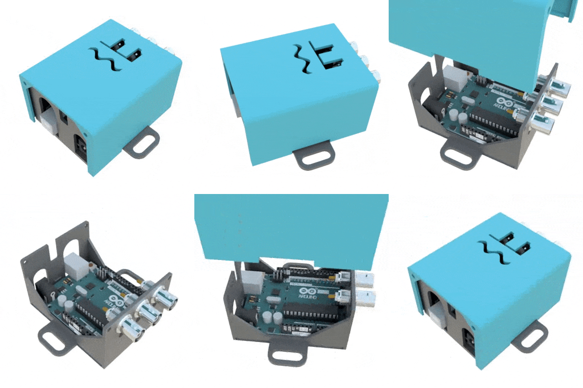
SignalBuddy is an easy-to-make, easy-to-use signal generator for scientific applications. Making friends is hard, but making SignalBuddy is easy. All you need is an Arduino Uno! SignalBuddy replaces more complicated and (much) more expensive signal generators in laboratory settings where one millisecond resolution is sufficient. SignalBuddy generates digital or true analog signals (sine waves, step functions, and pulse trains), can be controlled with an intuitive serial monitor interface, and looks fabulous in an optional 3D printed enclosure.

To get SignalBuddy working, all you need to do is install the SignalBuddy.ino Arduino code provided on their github, and follow the step-by-step instructions on github to get the Arduino programmed up for your specific experimental needs. SignalBuddy can be used for numerous lab purposes, including creating pulse trains for optogenetic light stimulation, microstimulation, electrophysiology, or for programming up stimuli for behavioral paradigms.

Additionally, their hackaday site provides the instructions for 3D printing an enclosure to house the Arduino inside using just two .stl files.


For more information, check out the SignalBuddy github repository here.

You can also get further details on the SignalBuddy Hackaday.io page here.


Fun Fact: This group also developed KineMouse Wheel, a project previously posted on OpenBehavior and is now being used in numerous labs! Cheers to another great open-source project from Richard Warren and the Sawtell lab!

3D Printed Headstage Implant

June 6, 2019

Richard Pinnell from Ulrich Hofmann’s lab has three publications centered around open-source and 3D printed methods for headstage implant protection and portable / waterproof DBS and EEG to pair with water maze activity. We share details on the three studies below:


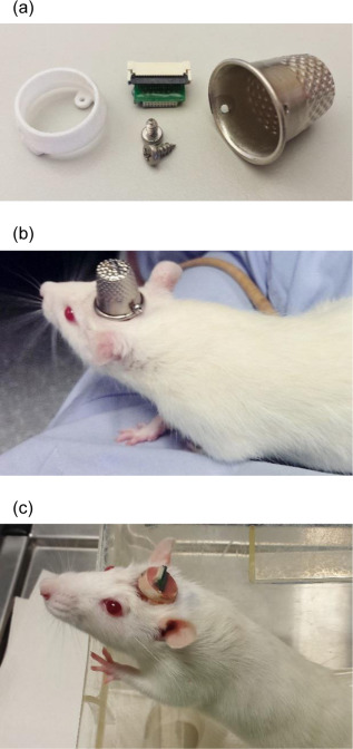
Most researchers opt to single-house rodents after rodents have undergone surgery. This helps the wound heal and prevent any issues with damage to the implant. However, there is substantial benefits to socially-housing rodents, as social isolation can create stressors for them. As a way to continue to socially-house rats, Pinnell et al. (2016a) created a novel 3D-printed headstage socket to surround an electrode connector. Rats were able to successfully be pair housed with these implants and their protective caps.

The polyamide headcap socket itself is 3D printed, and a stainless steel thimble can be screwed into it. The thimble can be removed by being unscrewed to reveal the electrode connector. This implant allows both for increased well-being of the rodent post-surgery, but also has additional benefits in that it can prevent any damage to the electrode implant during experiments and keeps the electrode implant clean as well.

The 3D printed headcap was used in a second study (Pinnell et al., 2016b) for wireless EEG recording in rats during a water maze task. The headstage socket housed the PCB electrode connector and the waterproof wireless system was attached. In this setup, during normal housing conditions, this waterproof attachment was replaced with a standard 18×9 mm stainless-steel sewing thimble, which contained 1.2 mm holes drilled at either end for attachment to the headstage socket. A PCB connector was manufactured to fit inside the socket, and contains an 18-pin zif connector, two DIP connectors, and an 18-pin Omnetics electrode connector for providing an interface between the implanted electrodes and the wireless recording system.

Finally, the implant was utilized in a third study (Pinnell et al., 2018) where the same group created a miniaturized, programmable deep-brain stimulator for use in a water maze. A portable deep brain stimulation (DBS) device was created through using a PCB design, and this was paired with the 3D printed device. The 3D printed headcap was modified from its use in Pinnell et al., 2016a to completely cover the implant and protect the PCB. The device, its battery, and housing weighs 2.7 g, and offers protection from both the environment and from other rats, and can be used in DBS studies during behavior in a water maze.

The portable stimulator, 3D printed cap .stl files, and more files from the publications can be found on https://figshare.com/s/31122e0263c47fa5dabd.


Pinnell, R. C., Almajidy, R. K., & Hofmann, U. G. (2016a). Versatile 3D-printed headstage implant for group housing of rodents. Journal of neuroscience methods, 257, 134-138.

Pinnell, R. C., Almajidy, R. K., Kirch, R. D., Cassel, J. C., & Hofmann, U. G. (2016b). A wireless EEG recording method for rat use inside the water maze. PloS one, 11(2), e0147730.

Actifield

March 21, 2019

Victor Wumbor-Apin Kumbol and colleagues have developed and shared Actifield, an automated open-source actimeter for rodents, in a recent HardwareX publication.


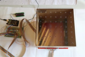
Measuring locomotor activity can be a useful readout for understanding effects of a number of experimental manipulations related to neuroscience research. Commercially available locomotor activity recording devices can be cost-prohibitive and often lack the ability to be customized to fit a specific lab’s needs. Kumbol et al. offer an open-source alternative that utilizes infrared motion detection and an arduino to record activity in a variety of chamber set ups. A full list of build materials, links to 3D-print and laser-cut files, and assembly instructions are available in their publication.

Read more from HardwareX!


TRIO Platform

December 12, 2018

Vladislav Voziyanov and colleagues have developed and shared the TRIO Platform, a low-profile in vivo imaging support and restraint system for mice.


In vivo optical imaging methods are common tools for understanding neural function in mice. This technique is often performed in head-fixed,  anesthetized animals, which requires monitoring of anesthesia level and body temperature while stabilizing the head. Fitting each of the components necessary for these experiments on a standard microscope stage can be rather difficult. Voziyanov and colleagues have shared their design for the TRIO (Three-In-One) Platform. This system is compact and  provides sturdy head fixation, a gas anesthesia mask, and warm water bed. While the design is compact enough to work with a variety of microscope stages, the use of 3D printed components makes this design customizable.

https://www.frontiersin.org/files/Articles/184541/fnins-10-00169-HTML/image_m/fnins-10-00169-g004.jpg

Read more about the TRIO Platform in Frontiers in Neuroscience!

The design files and list of commercially available build components are provided here.


PriED: An Open Source 3-D Printed Modular Micro-Drive System for Acute Neurophysiology

August 1, 2018

In a 2014 PLoS ONE article, Shaun R. Patel and colleagues share their design for PriED, an easy to assemble modular micro-drive system for acute primate neurophysiology.


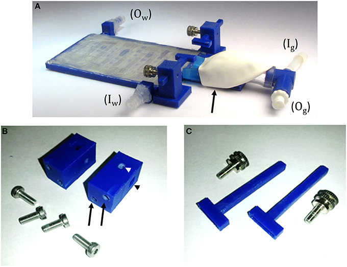
Electrode micro-drives are a great tool that allow for independent positioning of multiple electrodes in primate neurophysiology, however, commercially available micro-drives are often expensive. Printed Electronic Device (PriED) is designed to advance existing micro-drive technology while staying inexpensive and requiring minimal skill and effort to assemble. The device combines 3D printed parts and affordable, commercially available steel and brass components which can then be controlled manually, or automatically with the addition of an optional motor. Using 3D printing technology researchers have the flexibility to be able to modify part designs and create custom solutions to specific recording needs. A public repository of drive designs has been made available where researchers can download PriED components to print for assembly. Additionally, researchers can upload modified designs with annotations for others to use. PriED is an innovative, inexpensive, and user friendly micro-drive solution for flexible multi-site cortical and subcortical recordings in non-human primates.

Read more here!

Or check out the repository here!


Syringe Pump – Pearce Research Group

In their 2014 paper “Open-Source Syringe Pump Library,” Bas Wijnen, Emily Hunt, Gerald Anzalone, and Joshua Pearce detail an open-source syringe pump apparatus developed in their lab, as well as, validate the performance of the device. The authors write, “This syringe pump was designed using freely available open-source computer aided design (CAD) software and manufactured using an open-source RepRap 3-D printer and readily available parts. The design, bill of materials and assembly instructions are globally available to anyone wishing to use them on the Open-source syringe pump Approdepia page… The cost of the entire system, including the controller and web-based control interface, is on the order of 5% or less than one would expect to pay for a commercial syringe pump having similar performance. The design should suit the needs of a given research activity requiring a syringe pump including carefully controlled dosing of reagents, pharmaceuticals, and delivery of viscous 3-D printer media among other applications.”

Pearce Research group also provides an Open Source Lab page dedicated to low-cost, open-source lab hardware.


Wijnen, Bas; Hunt, Emily; Anzalone, Gerald; Pearce, Joshua (2014). Open-Source Syringe Pump Library. PLoS ONE, 9(9), e107216.