Tag: Implant

3D Printed Headcap and Microdrive

SEPTEMBER 26, 2019

In their 2015 Journal of Neurophysiology article, the Paré Lab at the Center for Molecular and Behavioral Neuroscience at Rutgers University describe their novel head-cap and microdrive design for chronic multi-electrode recordings in rats through the use of 3D printing technology and highlight the impact of 3D printing technology on neurophysiology:


There is a need for microdrives and head-caps that can accommodate different recording configurations. Many investigators implant multiple individual drives aiming to record from numerous areas. However, this extends surgery time, impairs animal recovery, and complicates experiments. Other strategies rely on more expensive custom-machined drive assemblies that are specifically built for a particular set of regions, limiting their adaptability. Some proposed designs allow targeting of multiple regions, but recording sites must be within a few millimeters so are only suitable for mice and not for accessing areas of larger brains (like in rats, for example).

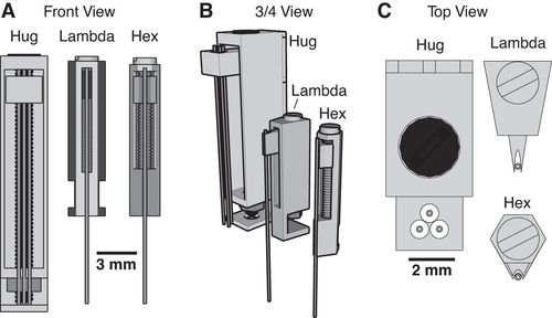
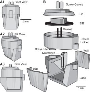
Utilizing 3D printing technology to create a novel design concept of microdrives and head-caps, this group’s design allows for recording of multiple brain regions in different configurations. In their article, the lab reviews the basic principles of 3D design and printing and introduce their approach to multisite recording, explaining how to construct the individual required components. The 3D printed head cap and electrode microdrive enables investigators to perform chronic multi-site recordings in rats. The head cap is composed of five components and there are three types of microdrives that can be used in different combinations or positions to study different targets. The different microdrive designs have different functionality including for extended driving depths, targeting of thin layers, and allowing many microdrives to be placed in a small area.

To show the viability of their new designs, the lab presents LFP recordings obtained throughout the cortico-hippocampal loop using 3D printed components. The lab suggests investigators modify their designs to best suit their research needs and give changeable versions of the three parts most important in modification. The investigators also provide a detailed explanation of the printing, assembly, and implantation of the head caps and microdrives. Finally, they indicate the ways 3D printing advancements can change how chronic implants are designed and used, notably 3D scanning and new material development.

For more information on the microdrive and headcap, see their paper’s Appendix, which has full instructions and advice on building these devices.


Headley, D. B., DeLucca, M. V., Haufler, D., & Paré, D. (2015). Incorporating 3D-printing technology in the design of head-caps and electrode drives for recording neurons in multiple brain regions. Journal of Neurophysiology, 113(7), 2721–2732. https://doi.org/10.1152/jn.00955.2014
HOPE

July 12, 2019

Sebastien Delcasso from the Graybiel lab at MIT published a method for developing a brain implant called “HOPE” for combining with optogenetics, pharmacology, and electrophysiology:


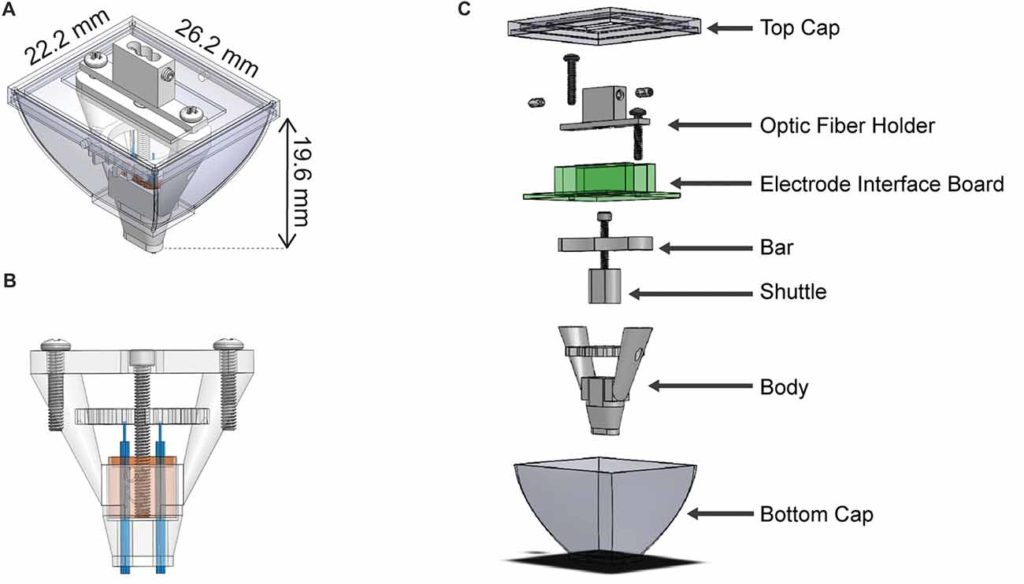
HOPE (hybrid-drive combining optogenetics, pharmacology, and electrophysiology) is a method that simplifies the construction of a drivable and multi-task recording implant. HOPE is a new type of implant that can support up to 16 tetrodes, and allows for recordings of two different brain areas in a mouse at the same time, along with simultaneous optogenetic or pharmacological manipulation. The HOPE implants are open-source and can be recreated in CAD software and subsequently 3D printed, drastically lowering the cost of an electrophysiological implant. Additionally, instead of waiting months for a custom-made implant, these can be printed within a few hours.

The manuscript provides detailed instructions on constructing the implant, and allows for users to individually modify it for their own needs (and can be modified to be used in rats or non-human primates). Additionally, HOPE is meant to be used in experiments with paired electrophysiological experiments with either optogenetic or pharmacological manipulations, which will inevitably open the door to many more experiments. The implant is intended for microdrive recordings, and the actual implant is only made up of six 3D printed parts, an electrode interface board (EIB), and five screws.

The authors validate the implant by first successfully recording striatal neurons, using transgenic PV-Cre mice to optogenetically inhibit parvalbumin interneurons, and then using muscimol infused into the striatum in a head-fixed mouse preparation. HOPE is a novel open-source neural implant that can be paired with multiple methods (recordings, optogenetics, and pharmacology) to help in manipulating and subsequently recording brain activity.


More details of their implant can be found on their project site and on the project GitHub.


Delcasso, S., Denagamage, S., Britton, Z., & Graybiel, A. M. (2018). HOPE: Hybrid-Drive Combining Optogenetics, Pharmacology and Electrophysiology. Frontiers in neural circuits, 12, 41.


3D Printed Headstage Implant

June 6, 2019

Richard Pinnell from Ulrich Hofmann’s lab has three publications centered around open-source and 3D printed methods for headstage implant protection and portable / waterproof DBS and EEG to pair with water maze activity. We share details on the three studies below:


Most researchers opt to single-house rodents after rodents have undergone surgery. This helps the wound heal and prevent any issues with damage to the implant. However, there is substantial benefits to socially-housing rodents, as social isolation can create stressors for them. As a way to continue to socially-house rats, Pinnell et al. (2016a) created a novel 3D-printed headstage socket to surround an electrode connector. Rats were able to successfully be pair housed with these implants and their protective caps.

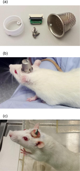
The polyamide headcap socket itself is 3D printed, and a stainless steel thimble can be screwed into it. The thimble can be removed by being unscrewed to reveal the electrode connector. This implant allows both for increased well-being of the rodent post-surgery, but also has additional benefits in that it can prevent any damage to the electrode implant during experiments and keeps the electrode implant clean as well.

The 3D printed headcap was used in a second study (Pinnell et al., 2016b) for wireless EEG recording in rats during a water maze task. The headstage socket housed the PCB electrode connector and the waterproof wireless system was attached. In this setup, during normal housing conditions, this waterproof attachment was replaced with a standard 18×9 mm stainless-steel sewing thimble, which contained 1.2 mm holes drilled at either end for attachment to the headstage socket. A PCB connector was manufactured to fit inside the socket, and contains an 18-pin zif connector, two DIP connectors, and an 18-pin Omnetics electrode connector for providing an interface between the implanted electrodes and the wireless recording system.

Finally, the implant was utilized in a third study (Pinnell et al., 2018) where the same group created a miniaturized, programmable deep-brain stimulator for use in a water maze. A portable deep brain stimulation (DBS) device was created through using a PCB design, and this was paired with the 3D printed device. The 3D printed headcap was modified from its use in Pinnell et al., 2016a to completely cover the implant and protect the PCB. The device, its battery, and housing weighs 2.7 g, and offers protection from both the environment and from other rats, and can be used in DBS studies during behavior in a water maze.

The portable stimulator, 3D printed cap .stl files, and more files from the publications can be found on https://figshare.com/s/31122e0263c47fa5dabd.


Pinnell, R. C., Almajidy, R. K., & Hofmann, U. G. (2016a). Versatile 3D-printed headstage implant for group housing of rodents. Journal of neuroscience methods, 257, 134-138.

Pinnell, R. C., Almajidy, R. K., Kirch, R. D., Cassel, J. C., & Hofmann, U. G. (2016b). A wireless EEG recording method for rat use inside the water maze. PloS one, 11(2), e0147730.