Category: Neural Recording

3D Printed Headcap and Microdrive

SEPTEMBER 26, 2019

In their 2015 Journal of Neurophysiology article, the Paré Lab at the Center for Molecular and Behavioral Neuroscience at Rutgers University describe their novel head-cap and microdrive design for chronic multi-electrode recordings in rats through the use of 3D printing technology and highlight the impact of 3D printing technology on neurophysiology:


There is a need for microdrives and head-caps that can accommodate different recording configurations. Many investigators implant multiple individual drives aiming to record from numerous areas. However, this extends surgery time, impairs animal recovery, and complicates experiments. Other strategies rely on more expensive custom-machined drive assemblies that are specifically built for a particular set of regions, limiting their adaptability. Some proposed designs allow targeting of multiple regions, but recording sites must be within a few millimeters so are only suitable for mice and not for accessing areas of larger brains (like in rats, for example).

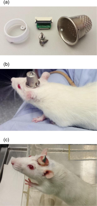
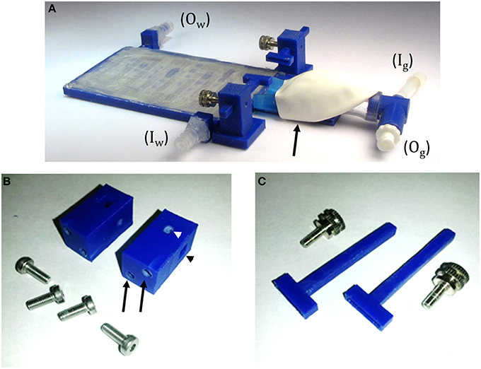
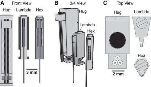
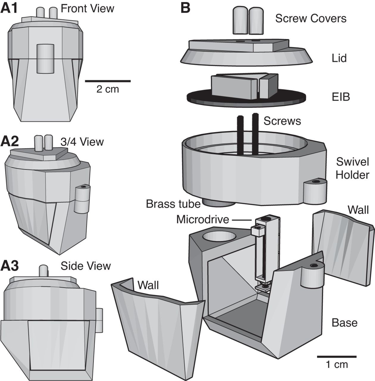
Utilizing 3D printing technology to create a novel design concept of microdrives and head-caps, this group’s design allows for recording of multiple brain regions in different configurations. In their article, the lab reviews the basic principles of 3D design and printing and introduce their approach to multisite recording, explaining how to construct the individual required components. The 3D printed head cap and electrode microdrive enables investigators to perform chronic multi-site recordings in rats. The head cap is composed of five components and there are three types of microdrives that can be used in different combinations or positions to study different targets. The different microdrive designs have different functionality including for extended driving depths, targeting of thin layers, and allowing many microdrives to be placed in a small area.

To show the viability of their new designs, the lab presents LFP recordings obtained throughout the cortico-hippocampal loop using 3D printed components. The lab suggests investigators modify their designs to best suit their research needs and give changeable versions of the three parts most important in modification. The investigators also provide a detailed explanation of the printing, assembly, and implantation of the head caps and microdrives. Finally, they indicate the ways 3D printing advancements can change how chronic implants are designed and used, notably 3D scanning and new material development.

For more information on the microdrive and headcap, see their paper’s Appendix, which has full instructions and advice on building these devices.


Headley, D. B., DeLucca, M. V., Haufler, D., & Paré, D. (2015). Incorporating 3D-printing technology in the design of head-caps and electrode drives for recording neurons in multiple brain regions. Journal of Neurophysiology, 113(7), 2721–2732. https://doi.org/10.1152/jn.00955.2014

SpikeGadgets

AUGUST 22, 2019

We’d like to highlight groups and companies that support an open-source framework to their software and/or hardware in behavioral neuroscience. One of these groups is SpikeGadgets, a company co-founded by Mattias Karlsson and Magnus Karlsson.


SpikeGadgets is a group of electrophysiologists and engineers who are working to develop neuroscience hardware and software tools. Their open-source software, Trodes, is a cross-platform software suite for neuroscience data acquisition and experimental control, which is made up of modules that communicate with a centralized GUI to visualize and save electrophysiological data. Trodes has a camera module and a StateScript module, which is a state-based scripting language that can be used to program behavioral tasks through using lights, levels, beam breaks, lasers, stimulation sources, audio, solenoids, etc. The camera module can be used to acquire video that can synchronize to neural recordings; the camera module can track the animal’s position in real-time or play it back after the experiment. The camera module can work with USB webcams or GigE cameras.

Paired with the Trodes software and StateScript language is the SpikeGadgets hardware that can be purchased on their website. The hardware is used for data acquisition (Main Control Unit, used for electrophysiology) and behavioral control (Environmental Control Unit).  SpikeGadgets also provides both Matlab and Python toolboxes on their site that can be used to analyze both behavioral and electrophysiological data. Trodes can be used on Windows, Linux, or Mac, and there are step-by-step instructions for how to install and use Trodes on the group’s bitbucket page.

Spikegadgets mission is “to develop the most advanced neuroscience tools on the market, while preserving ease of use and science-driven customization.”


For more information on SpikeGadgets or to download or purchase their software or hardware, check out their website here.

There is additional documentation on their BitBucket Wiki, with a user manual, instructions for installation, and FAQ.

Check out their entire list of collaborators, contributors, and developers here.

HOPE

July 12, 2019

Sebastien Delcasso from the Graybiel lab at MIT published a method for developing a brain implant called “HOPE” for combining with optogenetics, pharmacology, and electrophysiology:


HOPE (hybrid-drive combining optogenetics, pharmacology, and electrophysiology) is a method that simplifies the construction of a drivable and multi-task recording implant. HOPE is a new type of implant that can support up to 16 tetrodes, and allows for recordings of two different brain areas in a mouse at the same time, along with simultaneous optogenetic or pharmacological manipulation. The HOPE implants are open-source and can be recreated in CAD software and subsequently 3D printed, drastically lowering the cost of an electrophysiological implant. Additionally, instead of waiting months for a custom-made implant, these can be printed within a few hours.

The manuscript provides detailed instructions on constructing the implant, and allows for users to individually modify it for their own needs (and can be modified to be used in rats or non-human primates). Additionally, HOPE is meant to be used in experiments with paired electrophysiological experiments with either optogenetic or pharmacological manipulations, which will inevitably open the door to many more experiments. The implant is intended for microdrive recordings, and the actual implant is only made up of six 3D printed parts, an electrode interface board (EIB), and five screws.

The authors validate the implant by first successfully recording striatal neurons, using transgenic PV-Cre mice to optogenetically inhibit parvalbumin interneurons, and then using muscimol infused into the striatum in a head-fixed mouse preparation. HOPE is a novel open-source neural implant that can be paired with multiple methods (recordings, optogenetics, and pharmacology) to help in manipulating and subsequently recording brain activity.


More details of their implant can be found on their project site and on the project GitHub.


Delcasso, S., Denagamage, S., Britton, Z., & Graybiel, A. M. (2018). HOPE: Hybrid-Drive Combining Optogenetics, Pharmacology and Electrophysiology. Frontiers in neural circuits, 12, 41.


3D Printed Headstage Implant

June 6, 2019

Richard Pinnell from Ulrich Hofmann’s lab has three publications centered around open-source and 3D printed methods for headstage implant protection and portable / waterproof DBS and EEG to pair with water maze activity. We share details on the three studies below:


Most researchers opt to single-house rodents after rodents have undergone surgery. This helps the wound heal and prevent any issues with damage to the implant. However, there is substantial benefits to socially-housing rodents, as social isolation can create stressors for them. As a way to continue to socially-house rats, Pinnell et al. (2016a) created a novel 3D-printed headstage socket to surround an electrode connector. Rats were able to successfully be pair housed with these implants and their protective caps.

The polyamide headcap socket itself is 3D printed, and a stainless steel thimble can be screwed into it. The thimble can be removed by being unscrewed to reveal the electrode connector. This implant allows both for increased well-being of the rodent post-surgery, but also has additional benefits in that it can prevent any damage to the electrode implant during experiments and keeps the electrode implant clean as well.

The 3D printed headcap was used in a second study (Pinnell et al., 2016b) for wireless EEG recording in rats during a water maze task. The headstage socket housed the PCB electrode connector and the waterproof wireless system was attached. In this setup, during normal housing conditions, this waterproof attachment was replaced with a standard 18×9 mm stainless-steel sewing thimble, which contained 1.2 mm holes drilled at either end for attachment to the headstage socket. A PCB connector was manufactured to fit inside the socket, and contains an 18-pin zif connector, two DIP connectors, and an 18-pin Omnetics electrode connector for providing an interface between the implanted electrodes and the wireless recording system.

Finally, the implant was utilized in a third study (Pinnell et al., 2018) where the same group created a miniaturized, programmable deep-brain stimulator for use in a water maze. A portable deep brain stimulation (DBS) device was created through using a PCB design, and this was paired with the 3D printed device. The 3D printed headcap was modified from its use in Pinnell et al., 2016a to completely cover the implant and protect the PCB. The device, its battery, and housing weighs 2.7 g, and offers protection from both the environment and from other rats, and can be used in DBS studies during behavior in a water maze.

The portable stimulator, 3D printed cap .stl files, and more files from the publications can be found on https://figshare.com/s/31122e0263c47fa5dabd.


Pinnell, R. C., Almajidy, R. K., & Hofmann, U. G. (2016a). Versatile 3D-printed headstage implant for group housing of rodents. Journal of neuroscience methods, 257, 134-138.

Pinnell, R. C., Almajidy, R. K., Kirch, R. D., Cassel, J. C., & Hofmann, U. G. (2016b). A wireless EEG recording method for rat use inside the water maze. PloS one, 11(2), e0147730.

Telemetry System for Recording EEG

March 29, 2019

In a 2011 Journal of Neuroscience Methods article, Pishan Chang and colleagues shared their design for an open-source, novel telemetry system for recording EEG in small animals.


EEG monitoring in freely-behaving small animals is a useful technique for observing natural fluctuations in neural activity over time. Monitoring frequencies above 80 Hz continuously over a period of weeks can be a challenge. Chang et al. have shared their design for a system that combines an implantable telemetric sensor, radio-frequency transmission, and an open-source data acquisition software to collect EEG data over a span of up to 8 weeks. Various modifications to the system  have increased the longevity of the device and reduced transmission noise to provide continuous and reliable data. Schematics of the device, transmission system, and validation results in a population of epileptic rodents are available in their publication.


Learn more from the Journal of Neuroscience Methods!


Craniobot

March 13, 2019

Suhasa Kodandaramaiah from the University of Minnesota, Twin Cities, has shared the following about Craniobot, a computer numerical controlled robot for cranial microsurgeries.


The palette of tools available for neuroscientists to measure and manipulate the brain during behavioral experiments has greatly expanded in the previous decade. In many cases, using these tools requires removing sections of the skull to access the brain. The procedure to remove the sub-millimeter thick mouse skull precisely without damaging the underlying brain can be technically challenging and often takes significant skill and practice. This presents a potential obstacle for neuroscience labs wishing to adopt these technologies in their research. To overcome this challenge, a team at the University of Minnesota led by Mathew Rynes and Leila Ghanbari (equal contribution) created the ‘Craniobot,’ a cranial microsurgery platform that combines automated skull surface profiling with a computer numerical controlled (CNC) milling machine to perform a variety of cranial microsurgical procedures on mice. The Craniobot can be built from off-the-shelf components for a little over $1000 and the team has demonstrated its capability to perform small to large craniotomies, skull thinning procedures and for drilling pilot holes for installing bone anchor screws.

Read more about the Craniobot here. Software package for controlling the craniobot can be found on Github.


CAVE

In a recent article, Jennifer Tegtmeier and colleagues have shared CAVE: an open-source tool in MATLAB for combined analysis of head-mounted calcium imaging and behavior.


Calcium imaging is spreading through the neuroscience field like melted butter on hot toast. Like other imaging techniques, the data collected with calcium imaging is large and complex. CAVE (Calcium ActiVity Explorer) aims to analyze imaging data from head-mounted microscopes simultaneously with behavioral data. Tegtmeier et al. developed this software in MATLAB with a bundle of unique algorithms to specifically analyze single-photon imaging data, which can then be correlated to behavioral data. A streamlined workflow is available for novice users, with more advanced options available for advanced users. The code is available for download from GitHub.

Read more from Frontiers in Neuroscience, or check it out directly from GitHub.


Mousecam

February 6, 2019

Arne Meyer and colleagues recently shared their design and implementation of a head-mounted camera system for capturing detailed behavior in freely moving mice.


Video monitoring of animals can give great insight to behaviors. Most video monitoring systems to collect precise behavioral data require fixed position cameras and stationary animals, which can limit observation of natural behaviors. To address this, Meyer et al. developed a system which combines a lightweight head-mounted camera and head-movement sensors to detect behaviors in mice. The system, built using commercially available and 3D printed parts, can be used to monitor a variety of subtle behaviors including eye position, whisking, and ear movements in unrestrained animals. Furthermore, this device can be mounted in combination with neural implants for recording brain activity.

Read more here! You can also check out their github here. Documentation and files are also available on OpenEphys here.


CaImAn

January 23, 2019

Hot off the press in eLife, Andrea Giovannucci and colleagues have shared their open-source software library, CaImAn, for one and two-photon Calcium Imaging data Analysis.


In vivo calcium imaging has gained popularity in recent years for its ability to record large quantities of neural activity from multiple brain areas over extended time periods. With advanced tools for recording and collecting data comes large quantities of data. With large datasets comes a need for streamlined ways to analyze it. Giovannucci and colleagues have developed and shared a toolbox for analyzing complex calcium imaging datasets. CaImAn, developed in the open-source Python language (with optional implementation in MATLAB), is designed to correct for motion, estimate spikes, detect new neurons, and assess neuronal activity and locations in a given timeframe. The software can be used on pre-recorded data or can also enabled for real-time analysis. CaImAn is available to download with examples from GitHub, and more information can be obtained through reading the aforementioned manuscript.

Check out GitHub, or the article from eLife!


TRIO Platform

December 12, 2018

Vladislav Voziyanov and colleagues have developed and shared the TRIO Platform, a low-profile in vivo imaging support and restraint system for mice.


In vivo optical imaging methods are common tools for understanding neural function in mice. This technique is often performed in head-fixed,  anesthetized animals, which requires monitoring of anesthesia level and body temperature while stabilizing the head. Fitting each of the components necessary for these experiments on a standard microscope stage can be rather difficult. Voziyanov and colleagues have shared their design for the TRIO (Three-In-One) Platform. This system is compact and  provides sturdy head fixation, a gas anesthesia mask, and warm water bed. While the design is compact enough to work with a variety of microscope stages, the use of 3D printed components makes this design customizable.

https://www.frontiersin.org/files/Articles/184541/fnins-10-00169-HTML/image_m/fnins-10-00169-g004.jpg

Read more about the TRIO Platform in Frontiers in Neuroscience!

The design files and list of commercially available build components are provided here.